|
HIP DYSPLASIA IN DOGS A GUIDE FOR DOG
OWNERS BY JOHN FOSTER
|
|
|
DEVELOPMENTAL DEMANDS It is argued that dogs are not
barn with hips affected by dysplasia (unlikehumans suffering
from the disease). Hip modelling, otherwise termed development,
can worsen with the passing of time, most particularly during
the rapid growth phase between 14 and 26 weeks of age. Unwelcome
changes in anatomic relationships within the joint start in
early puppy hood with first usage and continue through into
young adult hood. Wear and tearfrom exercise of the distorted
joint is followed by varying amounts of inflammation and degeneration
resulting in remodelling change.
|
| |
 |
THE BVA/KC HD SCHEME All radiographs submitted to
the BVA/KC Hip Dysplasia Scheme are assessedby means of scoring.
The hip score is the sum of the points awarded for each of
nine radiographic features of both hip joints. The lower the
score the less the degree of hip dysplasia present. The minimum
(best) score for each hip iszero and the maximum (worst) is
53, giving a range for the total of 0 to 106. The average
score of the breed, or the 'breed mean score', is calculated
from all the scores recorded for a given breed and is shown
alongside its range there by giving a representation of the
overall hip status of the breed. All breeders wishing to try
to control HD should breed only from animals with hipscores
well below the breed mean score.Sires (fathers) to be bred
from should only be ones whose progeny (offspring) have achieved
consistently low scores. The same selection procedure shouldbe
used for bitches for breeding, since the use of animals with
higher than ideal scores may make the risk of producing offspring
with high scores much greater. This circumstance is not only
disappointing and potentially costly interms of compromised
breeding plans, but may lead to subsequent civil courtaction.
|
|
For the hip scoring scheme to be meaningful and successful
in the attempt to control this serious disease it is important
that all radiographs taken under the scheme are submitted
for scoring, whatever the apparent state of the hips, in order
that the information gathered is as relevant as possible.
It is only by this means that proper conclusions may be drawn
by the scheme's statistitians, geneticists and veterinary
advisers. It is not hard to understand why things happen so
quickly and how critical awhole series of factors in the dog's
life are, when realising some animals treble their size and
weight during a three- month period of adolescence. Getting
all the many nutritional needs in the right quantity, to the
right placeand at the right time requires a mastery of logistics.
However, it has to be realised that this apparent basic requirement
overlays the parts played by inheritance and other factors,
for example the type of exercise taken and the degree of body
weight.
|
| |
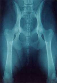
| SIGNS, APPEARANCE AND PATHOLOGY Hip dysplasia,
because it can be made up of a picture of joint looseness, new
bone formation or bone loss and inflammation and pain, can show
up in arange of signs from apparent soundness through lameness
to degrees of exercise intolerance. Combine these findings with
the fact that some breeds and some individuals are more stoical
than others and there is no predicting, just by looking from
the outside, to what degree a particular dog has or hasn't got
HD. More reliable is the clinical examination which is likely
to reveallimitation of movement of the affected hip, probably
reduction in muscle mass of the limb and some degree of pain.
Remember, a dog with HD in the normal course of life does not
show discomfort by, say, yelping, mainly because the pain is
likely to be continuous as opposed to sudden and unexpected.
The only way to assess properly the presence or relative absence
of HD is by radiography. This is an accurate photographic way
of showing the position ofthe ball of the joint in relation
to the socket and the presence and degree of any secondary changes. |
| |
|
|
| <<<
Structure & Function |
|
|
|